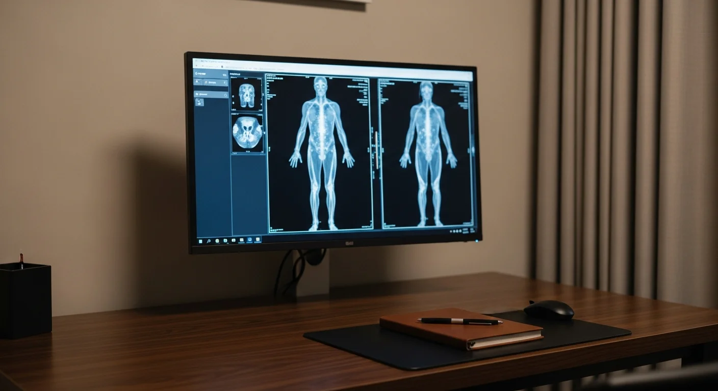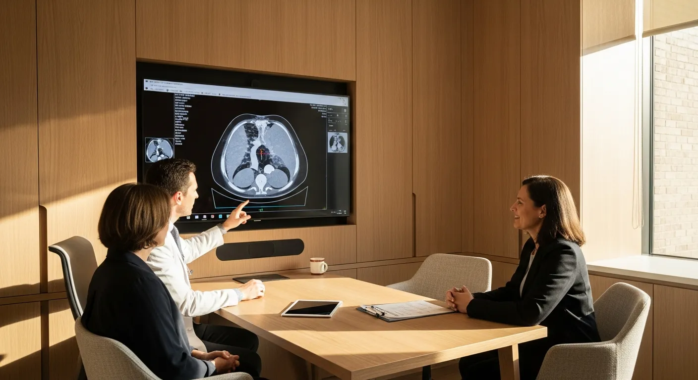
A chest CT resolves lung tissue in millimeters where a chest X-ray only shows large abnormalities, so it is the study for a persistent cough, a suspected blood clot, an abnormal X-ray, or interstitial lung disease. Fishtown Medicine matches the version of the scan to the question, since a nodule needs a non-contrast study, a suspected clot needs a timed CT angiogram, and scarring needs high-resolution slices. A standard chest CT delivers about 7 millisieverts against a natural background of roughly 3.1 per year.
TL;DR: A chest X-ray flattens the whole chest into one shadow, so anything smaller than a couple of centimeters, or anything hiding behind the heart, the ribs, or the diaphragm, can pass unnoticed. A CT takes hundreds of thin slices and reconstructs them, which is why it finds nodules of 3 or 4 mm that no plain film would have shown. That sensitivity is the reason to order it and the reason to be thoughtful about ordering it, because a large share of adults have small lung nodules that will never do anything, and finding one starts a follow-up process that carries its own worry. There are several versions of the study and they are not interchangeable: contrast timed to the arteries for a suspected clot, no contrast for nodules, high-resolution slices for scarring, and a low-dose protocol for screening. Which one you get should follow from the question being asked.
What does a chest CT show that an X-ray cannot?
A chest X-ray sends a single beam through your whole chest and records what reaches the detector, so everything from the skin on your back to the skin on your front is compressed into one image. Structures overlap, and the heart, the diaphragm, and the ribs all hide things behind them. It remains a good first study for pneumonia, fluid around the lung, a collapsed lung, and heart size, which is why it stays the front-line test for acute respiratory illness.
A CT rotates an X-ray source around you and reconstructs the data into cross-sections, typically about 1 mm thick. Nothing overlaps, because each slice shows only that plane. The practical consequence is a difference in what can be seen at all: a plain film generally needs an abnormality to reach roughly a centimeter or more before it separates from the background, while CT routinely resolves nodules of 2 to 3 mm and shows the fine architecture of lung tissue.
That is why a chest CT is the answer for a cough that has persisted for weeks without explanation, though a cough that long is more often walking pneumonia working its way through the city than anything a scan will name. The other clear indications are an abnormal or ambiguous chest X-ray, suspected lung cancer, staging of a known cancer, unexplained shortness of breath with a normal film, suspected bronchiectasis or interstitial lung disease, and any question about the mediastinum, the compartment in the middle of the chest holding the great vessels, lymph nodes, and thymus, which a plain film barely characterizes.
Which version of the scan do I need?
Asking for "a CT of the chest" is a bit like asking for "a blood test," since the protocol changes what the study can answer.
A non-contrast chest CT is the default for evaluating lung nodules, following a known nodule, and looking at lung tissue itself. Nodules are easier to measure and compare without contrast, and adding it would not improve the answer.
A CT pulmonary angiogram is a timed study where contrast is injected and images are captured while it fills the pulmonary arteries, which is how a blood clot in the lung is diagnosed. Timing is the whole trick, since scanning a few seconds early or late means the arteries are not opacified and the study cannot answer the question. This is an emergency evaluation, and sudden shortness of breath or pleuritic chest pain belongs in an emergency department rather than in an outpatient queue.
A contrast-enhanced chest CT is used when the question involves the mediastinum, a mass, lymph nodes, an infection that might have formed an abscess, or cancer staging, because contrast separates blood vessels from lymph nodes and shows how a mass takes up blood supply.
A high-resolution CT uses thinner slices and a reconstruction algorithm tuned for lung detail, and it is the study for suspected interstitial lung disease, pulmonary fibrosis, hypersensitivity pneumonitis, and bronchiectasis. A chronic cough with a smoking history or a suspicion of structural lung disease is a high-resolution question rather than a plain film question.
A low-dose CT is a stripped-down non-contrast protocol used for lung cancer screening in people who qualify by age and smoking history, delivering roughly a fifth of the radiation of a standard chest CT. That is its own pathway with its own eligibility rules, covered in the low-dose CT screening guide.
What about the radiation?
A standard chest CT delivers about 7 millisieverts of effective dose, against a chest X-ray at about 0.1 millisieverts and natural background radiation in the United States averaging roughly 3.1 millisieverts per year.1 A low-dose screening CT runs around 1.5 millisieverts.
So a chest CT is roughly 70 chest X-rays, or a bit over 2 years of background exposure. For a single study answering a question that matters, that is a reasonable trade and I would not hesitate. Where it deserves thought is in a person who is going to have many of them, which happens with nodule surveillance, with a cancer history, and with chronic lung disease. In those situations the conversation is about protocol and interval rather than about whether to image at all, and low-dose techniques can often be used for follow-up even outside the screening program.
Modern scanners have also reduced doses substantially over the past decade, so figures you may have read years ago tend to overstate what current equipment delivers.
How do I prepare, and what is the scan like?
For a non-contrast chest CT there is no preparation. You lie on a table, the table moves through a ring rather than a long tube, and the scan itself takes seconds. You will be asked to hold your breath for a few seconds at a time, and following that instruction matters more than people realize, since breathing during acquisition blurs the lung and can force a repeat.
Fishtown Medicine
A 90-minute conversation with Dr. Ash. A written plan you can follow.
For a study with contrast, expect a few extra steps. An intravenous line goes in, kidney function is usually checked beforehand in anyone over a certain age or with known kidney disease, and you should mention any previous reaction to contrast, any thyroid condition, and whether you take metformin. When the contrast goes in you will feel a spreading warmth through your chest and pelvis, and many people get a brief sensation that they have wet themselves. That is expected, it passes in under a minute, and technologists mention it ahead of time because it is alarming otherwise. Fasting for about 4 hours is typical for contrast studies.
The whole appointment usually runs 15 to 30 minutes, most of which is preparation rather than scanning. Unlike an MRI, a CT is quiet, open, and quick, so claustrophobia is rarely a barrier.
What happens if they find a nodule?
This is the part worth understanding before the scan rather than after, because a nodule is a common result and the word carries more weight than it should.
Small lung nodules are found frequently in adults, most are not cancer, and the great majority of the small ones are old scars from infections that resolved years ago. What determines the response is size, appearance, and your own risk, and the Fleischner Society guidelines lay out how those combine.2 The 2017 revision raised the size threshold below which no routine follow-up is recommended to 6 mm for solid nodules in low-risk people, and moved follow-up intervals from precise dates to ranges, both in recognition that the previous approach generated more scans and more anxiety than it prevented harm.
What that means in practice is that a report describing a 4 mm solid nodule in a person who never smoked frequently warrants no further imaging at all. A larger nodule, one with irregular margins, or one in a person with substantial smoking history gets a follow-up scan at an interval matched to the risk, and growth across scans is what changes the plan. Part-solid and ground-glass nodules follow different rules and are watched longer, because the slow-growing lesions behave differently from solid nodules.
The single most useful thing you can do is produce any prior chest imaging. A nodule that is unchanged across 3 years is a settled question, and the same nodule seen once is an open one. Old films from another health system are worth chasing down.
Guidance from the Clinic

How Fishtown Medicine approaches chest CT in Philadelphia
The protocol gets specified when the study is ordered rather than left to chance, because the wrong version of the scan means either a repeat or an answer to a question nobody asked. That means saying plainly whether contrast is needed, whether high-resolution slices are required, and what the clinical question is, so the radiologist is reading toward something.
For anyone who will be followed over time, continuity of facility matters as much as it does for ultrasound. Comparing a nodule across scans done on different machines with different slice thicknesses introduces uncertainty that a consistent setup avoids. Philadelphia has good independent imaging centers alongside the hospital systems, and for self-pay patients the price difference on a chest CT can be considerable.
Results come with a conversation. A chest CT report will list findings that have nothing to do with why it was ordered, from a small nodule to a bit of coronary calcium to a benign liver lesion caught at the bottom of the field, and each of those needs to be sorted into things that matter and things that do not.
Key Takeaways
- A chest CT resolves nodules of 2 to 3 mm where a chest X-ray generally needs about a centimeter, which is both the reason to order it and the reason to be deliberate about it.
- The protocol has to match the question: no contrast for nodules, timed contrast for a suspected clot, contrast for the mediastinum and staging, high-resolution slices for scarring, low-dose for screening.
- A standard chest CT is about 7 millisieverts, roughly 70 chest X-rays or a bit over 2 years of natural background.
- Small lung nodules are common and usually benign. Solid nodules under 6 mm in low-risk people often need no follow-up at all.
- Prior imaging is the most valuable and most overlooked test. A nodule unchanged over years answers a question a new scan cannot answer for months.
- Sudden shortness of breath or pleuritic chest pain is an emergency evaluation rather than an outpatient scan.
Related at Fishtown Medicine
- Lung Cancer Screening with Low-Dose CT - who qualifies and how the screening pathway works
- When You Need an X-Ray - what the chest film still does well
- Why We Choose an MRI - where MR fits and why it is rarely the chest answer
- Boutique Scans and Whole-Body MRI - the incidental finding problem in full
- When to Order Imaging - the framework behind choosing any scan
- Heart and Vascular Imaging - the cardiac studies that share the same scanner and the same field of view
- Chest Wall Pain - when pressing on the spot recreates the pain and imaging is not the answer
Scientific References
- Mettler FA Jr, Huda W, Yoshizumi TT, Mahesh M. "Effective doses in radiology and diagnostic nuclear medicine: a catalog." Radiology. 2008;248(1):254-263. PubMed
- MacMahon H, Naidich DP, Goo JM, et al. "Guidelines for Management of Incidental Pulmonary Nodules Detected on CT Images: From the Fleischner Society 2017." Radiology. 2017;284(1):228-243. PubMed

Frequently Asked Questions
Common Questions
Deep-Dive Questions
Ready when you are
The chat is our AI assistant, answering from our published guides. To talk it through with Dr. Ash himself, start with the intake.