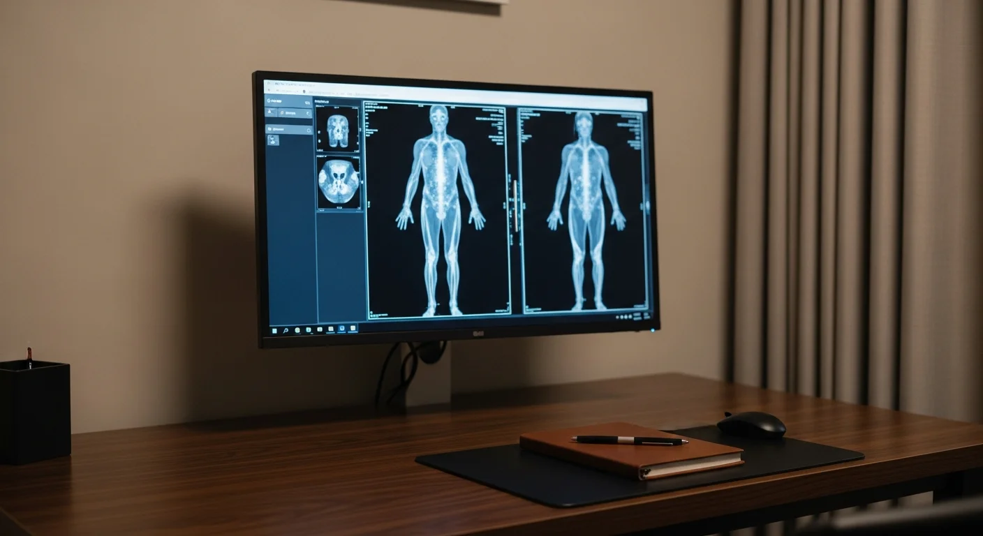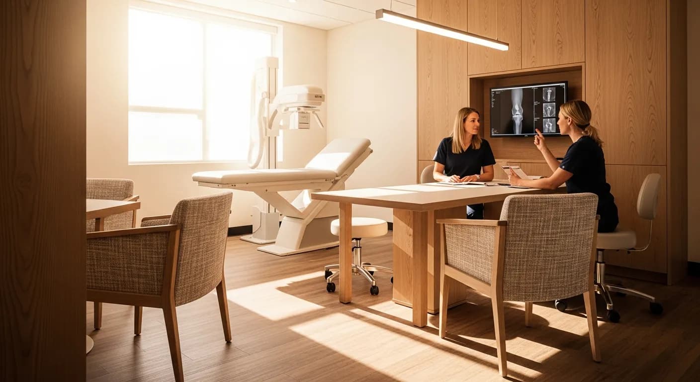
An X-ray answers questions about bone and alignment: fractures, arthritis, dislocations, and the position of hardware. It cannot see ligaments, cartilage detail, discs, or nerves, so a normal X-ray does not mean nothing is wrong. Fishtown Medicine orders plain films when the answer would change the plan, which usually means a defined injury, a red flag, or pain that has outlasted 4 weeks of conservative care. For most new joint and back pain without red flags, time and physical therapy outperform an early picture.
TL;DR: If something hurts and you are wondering whether you need a picture of it, the useful question is not whether an X-ray is available. It is whether the result would change what happens next. An X-ray is a bone test. It is very good at fractures, alignment, arthritis, and hardware, and it is close to blind for ligaments, cartilage surfaces, discs, and nerves, which means a normal film is common in people who are in a great deal of pain. Radiation for a limb is trivial, around 0.001 millisieverts, while a lumbar spine series runs closer to 1.5 millisieverts against a natural background of about 3.1 millisieverts a year. For most new back, neck, and joint pain with no red flags, the evidence favors 4 weeks of conservative care before any imaging, because early films do not improve outcomes and do lead to more procedures. The sections below are organized by body part, so skip to yours.
What can an X-ray show, and what does it miss?
An X-ray is a shadow. Dense tissue such as bone absorbs the beam and shows up white, air shows up black, and everything in between is a gradient of grey. That physics is the whole story of what the study can and cannot answer for you.
What it shows well is bone and the space between bones. A fracture line, a bone that has moved out of position, the narrowing of a joint space that tells you cartilage has thinned, the bone spurs of osteoarthritis, a dislocation, a foreign body such as glass or metal, and the position of a plate or screw someone put in years ago. In the chest it also shows air and fluid where they should not be, which is why it remains the first study for a cough with fever.
What it misses is most of what people assume it covers. Ligaments, tendons, cartilage surfaces, menisci, labrums, spinal discs, and nerves are all soft tissue, and they are close enough to each other in density that the film cannot separate them. A torn rotator cuff, a blown ACL, a herniated disc pressing on a nerve root, and a stress fracture in its first 2 weeks can all sit behind a film that gets read as normal. This is the part I want you to hold onto, because a normal X-ray is often heard as "nothing is wrong with you," and what it means is narrower than that: the bones look intact in the planes we photographed.
How much radiation is in an X-ray?
The dose depends enormously on the body part, and the spread is wider than most people expect. Using the standard catalog of effective doses, a limb such as a hand, wrist, elbow, foot, or knee runs about 0.001 millisieverts, a chest X-ray about 0.1 millisieverts, an abdominal film about 0.7 millisieverts, and a lumbar spine series about 1.5 millisieverts.1 Natural background radiation in the United States averages roughly 3.1 millisieverts a year, so an X-ray of your elbow is a rounding error against a week of simply being alive, while a lumbar spine series is closer to 6 months of background.
That range flips the usual intuition. People often worry about the wrist film and shrug at the back film, when the back film carries something like 1,500 times the dose. None of these numbers should frighten you away from a study that will answer a question. They are here so that the decision is made on whether the picture helps, which is the argument that matters, rather than on a vague sense that radiation is bad.
Why is waiting often the right first move?
For new back, neck, and most joint pain without red flags, the evidence has been consistent for a long time: imaging early does not make people better. A meta-analysis of 6 randomized trials covering 1,804 patients with low back pain found no meaningful difference between immediate lumbar imaging and usual care without imaging, for pain or for function, at 3 months and at 6 to 12 months.2 The American College of Physicians guideline reached the same place and recommends against routine imaging for nonspecific low back pain.3
The reason is not that we are saving money on your behalf. It is that scans of adults over 30 are full of findings that look alarming and mean very little. Disc bulges, degenerative changes, and small tears show up in large numbers of people who have no pain at all, so once a picture exists, both of us have to work out whether the thing on the film is the thing causing the symptom. That uncertainty pushes toward more tests and more procedures without making the pain better.
So in my practice the default for new pain with no red flags is a defined 4-week window of conservative care, which usually means physical therapy, activity modification, and something reasonable for pain, with a specific plan for what happens if it does not improve. The window matters as much as the waiting. Waiting without a follow-up date is just being left alone, and that is not what I mean.
What overrides the waiting is a red flag. A significant injury, an inability to bear weight, a history of cancer, fever with the pain, unexplained weight loss, a progressive loss of strength or sensation, or any change in bowel or bladder control moves you straight to imaging or to an emergency department. Bowel or bladder changes with back pain need care the same day.
Chest X-ray
A chest X-ray answers questions about the lungs, the pleural space, the heart's silhouette, and the bony thorax, and it is the first imaging study for most acute respiratory illness. The standard is 2 views, one from back to front and one from the side, because a single frontal view hides a surprising amount behind the heart and the diaphragm.
It is the right study when there is a cough with fever, shortness of breath, chest pain that might be pleuritic, a suspected pneumonia, or a concern about fluid around the lung. It also picks up a collapsed lung, an enlarged heart, and larger masses.
What it will not do is rule out a blood clot in the lung, which needs a CT angiogram, and it is a poor screening test for lung cancer. Tumors have to reach a fair size before they separate from the surrounding structures on a plain film, which is why lung cancer screening for people with significant smoking history uses low-dose CT rather than chest X-ray. If that applies to you, the low-dose CT screening guide is the piece to read. A chronic cough that has gone on for weeks with a smoking history or a suspicion of structural lung disease is also a CT question rather than a plain film question, and the chest CT guide covers which version of that scan answers which question. A cough that has dragged on for a month without fever is more often walking pneumonia than anything the film will show.
Neck and back X-rays
Spine films show alignment, vertebral body height, fractures, slippage of one vertebra on another, and the disc space narrowing and bone spurs that come with age. They do not show the disc itself, the spinal cord, or the nerve roots, so they cannot confirm or exclude a herniated disc or a pinched nerve.
For acute low back pain or isolated neck pain under 4 weeks with no red flags, the right answer is usually no imaging at all. Start physical therapy, stay as active as the pain allows, and set a date to reassess. If pain persists past 4 to 6 weeks despite a fair course of conservative care, a plain film becomes reasonable as a first look, particularly to check alignment and rule out a compression fracture.
The exceptions move fast. Meaningful trauma, a history of cancer, fever or a history of injection drug use, osteoporosis or long-term steroid use, new focal weakness, or saddle numbness and bowel or bladder changes all justify imaging now rather than in a month. Progressive weakness or any change in bowel or bladder control needs emergency care the same day rather than a scheduled visit.
MRI of the spine has a narrow set of jobs: surgical planning, a progressive neurologic deficit, or pain past 6 weeks that has fully failed physical therapy. Numbness alone, without weakness and without a failed course of therapy, is not by itself a reason to go to MRI first, because the finding rate in people without symptoms is high enough that the scan often adds confusion rather than clarity.
If a spine film comes back showing a wedge deformity or a compression fracture in someone younger without a serious injury behind it, that finding deserves a workup of its own rather than only a referral. New insufficiency fractures in a younger adult raise questions about thyroid function, parathyroid hormone, and vitamin D, and those labs should get run.
Shoulder X-ray
A shoulder series is 3 views, typically an AP, a true AP known as the Grashey view, and a scapular Y, and together they show the joint surfaces, the space under the acromion, arthritis, calcium deposits in the tendons, dislocation, and fractures.
Most shoulder pain that comes on without an injury is soft tissue, meaning rotator cuff tendinopathy, bursitis, or adhesive capsulitis, and none of those has a signature on a plain film. Even so I keep a low threshold for filming the shoulder, and the reason is a specific symptom: pain that wakes you from sleep. Night pain that pulls you out of a dead sleep is the one I take seriously, because it is one of the patterns that can point at the bone itself rather than the soft tissue around it. Suspected adhesive capsulitis is worth a film too, since the picture rules out the arthritis that would change the rehab plan.
A full-thickness rotator cuff tear is an MRI or ultrasound question, and the usual sequence is physical therapy first, then advanced imaging through orthopedics or sports medicine if the pain is severe, does not respond to therapy, or the exam suggests a complete tear.
Elbow X-ray
Elbow films are AP and lateral, and the lateral view carries most of the diagnostic weight because of a specific sign. Fat lies in the joint just outside the capsule, and when blood fills the joint after an injury it pushes that fat outward, making it visible. A posterior fat pad sign on a lateral elbow film in an adult means there is blood in the joint, and in practice that means a fracture is present even when no fracture line can be seen. In adults the usual culprit is a radial head fracture.
Fishtown Medicine
A 90-minute conversation with Dr. Ash. A written plan you can follow.
Film the elbow after a fall on an outstretched hand, after a direct blow, when the joint will not fully straighten or bend, or when there is swelling and point tenderness over bone. What the film will not explain is the far more common tennis elbow or golfer's elbow, which are tendon problems diagnosed by where it hurts and what movement reproduces it. A normal elbow X-ray in someone with lateral elbow pain and a positive resisted wrist extension test is the expected result rather than a puzzle.
Wrist and hand X-rays
Wrist and hand films show fractures, dislocations, arthritis, and the alignment of a healing injury. Standard wrist views are AP, lateral, and oblique, with dedicated scaphoid views added when the history calls for them.
The scaphoid is the reason this section exists. It is the small bone at the base of the thumb, in the hollow known as the anatomic snuffbox, and it is the classic fracture that hides on a first film. Pooled data put the prevalence of a radiographically occult scaphoid fracture at roughly 24% among wrists with a suspicious exam and normal initial X-rays.4 That matters more than the number suggests, because the blood supply to the scaphoid enters from one end, and a missed fracture can go on to lose its blood supply and fail to heal.
So the rule I follow is that a normal X-ray does not close the case when someone fell on an outstretched hand and is tender in the snuffbox. That wrist gets immobilized and re-imaged in 10 to 14 days, or gets an MRI sooner if waiting is not workable. Being splinted for 2 weeks for a wrist that turns out to be fine is a much better outcome than the alternative.
Hip and pelvis X-rays
The standard set is an AP view of the pelvis plus a lateral view of the affected hip, which shows joint space narrowing, arthritis, fractures, and the bone shapes behind impingement.
Where the pain lives changes the answer more than anything else here. Pain on the outside of the hip, over the bony point, with tenderness when you press it and pain lying on that side at night, is usually trochanteric bursitis or a tight iliotibial band. That picture does not need an early X-ray. It needs physical therapy, and it usually responds. Pain in the groin, deep and often described as a catch or a pinch, is more likely to be coming from inside the joint, and that is the presentation where a film earns its place.
For non-traumatic hip pain that has failed 4 weeks of conservative care, an AP pelvis and lateral hip is the right next step to look for arthritis or the cam and pincer shapes of impingement. A suspected labral tear is not a plain film question and not an ultrasound question, since a standard ultrasound cannot see inside the joint; that pathway runs through orthopedics, which will decide about an MRI arthrogram.
One warning worth carrying: in an older adult who falls and cannot bear weight, a normal hip X-ray does not exclude a fracture. Occult hip fractures are well described, and continued inability to walk after a fall justifies advanced imaging regardless of what the plain film showed.
Knee X-ray
Knee films are where a validated decision rule does most of the work. The Ottawa Knee Rule says an acute knee injury needs radiographs if any one of these is present: age 55 or older, tenderness at the head of the fibula, isolated tenderness of the kneecap, inability to bend the knee to 90 degrees, or inability to bear weight for 4 steps both at the time of injury and at the time of evaluation. In prospective validation across 1,096 patients it identified all 63 clinically important fractures, a sensitivity of 1.0, while cutting radiography use by about 28%.5
For knee pain that is not from a discrete injury, the question is arthritis, and the views have to be weight-bearing. A knee filmed lying down lets the joint space open up and can make significant cartilage loss look mild, so the useful series is a standing AP, a lateral, and a sunrise or Merchant view to see how the kneecap tracks in its groove.
MRI of the knee is not the next step for most knee pain. It belongs to people who feel the knee giving way and have failed physical therapy, or who had a clear traumatic event they can describe down to the moment. Ordering it earlier tends to find meniscal changes that are common at midlife and were probably there before the pain started.
Foot and ankle X-rays
Ankle and foot injuries have their own validated rule. In short, an ankle needs films when there is pain around the malleoli plus either bone tenderness along the back edge or tip of either malleolus, or an inability to take 4 steps. A foot needs films when there is midfoot pain plus either bone tenderness at the base of the 5th metatarsal or over the navicular, or an inability to take 4 steps. Applied with care, that rule keeps a great many sprained ankles out of radiology while still catching the fractures.
Two situations deserve extra care. A midfoot injury with swelling and bruising on the sole of the foot raises the question of a Lisfranc injury, and that one needs weight-bearing views, because the abnormal separation between the bones can close up when the foot is not loaded and hide on a relaxed film. And a stress fracture, the kind that builds over weeks in a runner or someone who increased their mileage quickly, is usually invisible on X-ray for the first 2 to 3 weeks, because there is nothing to see until healing bone starts to lay down. Point tenderness on a specific bone with a training history that fits is treated as a stress fracture even when the film is clear, with repeat imaging or MRI if it matters for timing.
Guidance from the Clinic

How Fishtown Medicine handles imaging in Philadelphia
Fishtown Medicine schedules imaging studies for patients rather than handing over an order and a phone number. For anyone paying cash or carrying a high deductible, that includes finding a good facility at a sane price, since the spread between a hospital-affiliated radiology department and an independent imaging center in the Philadelphia area can be several hundred dollars for the same study on comparable equipment.
Results come back to you with an explanation attached. A radiology report is written by one physician for another, and it is full of hedged phrasing and incidental findings that read as frightening out of context. The part that matters is what the finding means for you, whether it explains your symptom, and what happens next, and that is a conversation rather than a portal notification.
If you already have imaging from somewhere else, bring it. Prior studies are worth more than most people realize, because change over time is often the finding. A nodule that has been the same size for 4 years is a different situation from the same nodule seen once.
Key Takeaways
- An X-ray is a bone test. It answers fractures, alignment, arthritis, dislocation, and hardware position, and it cannot see ligaments, cartilage, discs, or nerves.
- A normal film means the bones look intact, not that nothing is wrong. Most joint pain comes from soft tissue that a plain film cannot show.
- For new back, neck, and joint pain with no red flags, 4 weeks of conservative care with a scheduled reassessment beats early imaging, which has not been shown to improve pain or function.
- Red flags override the waiting: significant trauma, inability to bear weight, cancer history, fever, unexplained weight loss, progressive weakness, or any bowel or bladder change.
- Radiation varies by 1,500-fold across studies. A limb film is about 0.001 millisieverts and a lumbar spine series about 1.5 millisieverts, against roughly 3.1 millisieverts of natural background per year.
- Two normal films that should not reassure you: a snuffbox-tender wrist after a fall, and an older adult who cannot bear weight after a fall.
Related at Fishtown Medicine
- Bone and Joint Imaging 101 - how plain films, ultrasound, and MRI fit together for musculoskeletal pain
- When to Order Imaging - the decision framework behind every scan we order
- Abdominal and Pelvic Imaging Guide - when the question is in the belly rather than the bones
- Lung Cancer Screening with Low-Dose CT - why chest X-ray is not a screening test
- Managing Labs and Imaging with Ease - how scheduling, cash pricing, and results-sharing work here
Scientific References
- Mettler FA Jr, Huda W, Yoshizumi TT, Mahesh M. "Effective doses in radiology and diagnostic nuclear medicine: a catalog." Radiology. 2008;248(1):254-263. PubMed
- Chou R, Fu R, Carrino JA, Deyo RA. "Imaging strategies for low-back pain: systematic review and meta-analysis." Lancet. 2009;373(9662):463-472. PubMed
- Qaseem A, Wilt TJ, McLean RM, Forciea MA. "Noninvasive Treatments for Acute, Subacute, and Chronic Low Back Pain: A Clinical Practice Guideline From the American College of Physicians." Annals of Internal Medicine. 2017;166(7):514-530. PubMed
- Ramadanov N, Marinova-Kichikova P, Dimitrov D, et al. "An Umbrella Review and Updated Meta-Analysis of Imaging Modalities in Occult Scaphoid and Hip and Femoral Fractures." Journal of Clinical Medicine. 2024;13(13):3769. Journal
- Stiell IG, Greenberg GH, Wells GA, et al. "Prospective validation of a decision rule for the use of radiography in acute knee injuries." JAMA. 1996;275(8):611-615. PubMed

Frequently Asked Questions
Common Questions
Deep-Dive Questions
Ready when you are
The chat is our AI assistant, answering from our published guides. To talk it through with Dr. Ash himself, start with the intake.