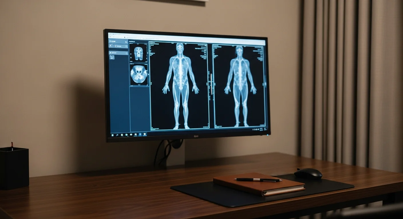
Ultrasound uses sound waves rather than radiation to image soft tissue as it moves, which makes it the first study for the thyroid, gallbladder, ovaries and uterus, testicles, and veins, and the safest choice in pregnancy. It cannot see through bone or gas, so it is poor for the lungs, the brain, and gas-filled bowel. Fishtown Medicine uses ultrasound when the question is about a cyst versus a mass, blood flow, or a superficial structure, and moves to CT or MRI when depth or bone is in the way.
TL;DR: Ultrasound is the study people worry about least and understand least. It sends sound into the body and listens for the echo, so there is no radiation, nothing to swallow in most cases, and no reason to avoid it during pregnancy. It is the first and often the final answer for the thyroid, the gallbladder, the ovaries and uterus, the testicles, and the veins of the leg, and it can watch things move, which no still image can do. Its limits are physical: sound will not travel through bone or air, so it cannot see the brain through an adult skull, cannot assess the lungs well, and struggles when gas-filled bowel lies between the probe and the target. It also depends more on the person holding the probe than any other study, which is why the facility matters. The sections below are organized by body part, so skip to yours.
What can an ultrasound show, and what is it blind to?
Ultrasound works by sending high-frequency sound into tissue and timing the echoes that come back. Different tissues reflect sound differently, and the machine assembles those echoes into a live picture. Two consequences follow from that, and they explain nearly everything about when this study is the right one.
The first is that ultrasound is superb at telling a fluid-filled cyst from a mass made of tissue. A cyst full of clear fluid lets sound pass straight through and looks black, while a nodule made of tissue scatters sound and looks grey and textured. That distinction is often the entire clinical question, whether it is a lump in the thyroid, a lump in the breast, or something on an ovary. Ultrasound also measures motion and flow, so with Doppler it can show blood moving through an artery, whether a vein is open or clotted, and how a heart valve behaves.
The second is that sound will not cross certain boundaries. Bone reflects almost all of it, and gas scatters almost all of it, so anything behind bone or behind bowel gas is hidden. That is why the lungs, the adult brain, and the deeper parts of the pancreas are poor ultrasound targets, and why a bowel full of gas can defeat an abdominal study on a given day.
The third consideration is human rather than physical. Ultrasound is acquired live by a person deciding where to point, how hard to press, and which images to save. A CT scan is the same study anywhere; an ultrasound is not. When the question is important, the facility and the technologist matter, and that is part of what I take on when I arrange a study for someone.
How do I prepare for an ultrasound?
Preparation depends on which study you are having, and getting it wrong is the most common reason a scan has to be repeated.
For an abdominal or right upper quadrant ultrasound looking at the gallbladder, liver, or pancreas, you fast for 6 to 8 hours beforehand. Food makes the gallbladder contract and empty, and a contracted gallbladder can hide stones that would be obvious in a full one. For a pelvic ultrasound done through the abdominal wall, the opposite applies: you drink water and arrive with a full bladder, because the bladder acts as a window that pushes bowel out of the way and lets sound reach the uterus and ovaries. For a transvaginal pelvic study, the bladder should be empty instead, and many appointments include both, which is why the instructions can sound contradictory. For the thyroid, the testicles, the veins of the leg, and most soft tissue studies, there is no preparation at all.
Wear something you can change out of easily, plan on gel that is warm at good facilities and startling at others, and expect the whole thing to take 20 to 45 minutes depending on the region.
Thyroid ultrasound
A thyroid ultrasound is the study for a lump in the neck, a gland that feels enlarged, or a nodule someone found incidentally on a scan done for another reason. It measures the gland, counts and sizes nodules, and describes each one in enough detail to estimate risk.
Thyroid nodules are common and overwhelmingly benign, which is the context that makes the rest of this section make sense. The job of the ultrasound is not to find nodules, since it will find them in a large share of adults, but to sort the ones that warrant a needle from the ones that warrant a repeat scan or nothing at all. The American College of Radiology system, TI-RADS, scores each nodule on composition, echogenicity, shape, margin, and the presence of bright foci, then pairs that score with the size to recommend biopsy, follow-up, or no further action.1
What I want you to take from that is the reason a report can say a nodule is 2 cm and still recommend leaving it alone, while another is 1 cm and needs a needle. Size alone does not decide. A nodule that is taller than it is wide, has an irregular margin, or contains punctate bright spots earns attention at a smaller size than a spongy, uniformly bright one does.
A thyroid ultrasound does not tell you how the gland is functioning. Whether you are hypothyroid or hyperthyroid is a blood question, and the two work together rather than substituting for each other.
Carotid ultrasound
Carotid ultrasound uses Doppler to look at the arteries running up either side of the neck. There are 2 separate reasons to do it, and confusing them leads to disappointment.
The first is stenosis, meaning narrowing that is already significant enough to threaten blood flow. That study is appropriate after a stroke or a transient ischemic attack, or when a bruit is heard over the artery, and it answers whether a surgical or procedural intervention should be considered.
The second is early plaque burden in someone with no symptoms at all, which is a prevention question rather than a surgical one. Measuring the thickness of the artery wall and looking for small plaques tells you whether atherosclerosis has begun, often decades before it would cause anything. That is a different study with a different purpose, and the carotid intima-media thickness guide covers how we use it alongside ApoB and Lp(a) to decide how proactive to be about a person's lipids.
Abdominal ultrasound
An abdominal or right upper quadrant ultrasound covers the gallbladder, bile ducts, liver, pancreas, spleen, kidneys, and the abdominal aorta, with fasting beforehand.
It is the first test for suspected gallstones, and it is very good at that job. Stones reflect sound strongly and cast a shadow behind them, and they move when you roll onto your side, which is the pattern a sonographer looks for. Pain in the right upper abdomen after fatty meals, sometimes radiating to the right shoulder blade, is the classic story that sends someone for this study.
It is also the first look at the liver, and it is where fatty liver is usually discovered, showing up as a gland that is brighter than the kidney next to it. That finding matters more than it used to, because metabolic dysfunction-associated fatty liver disease travels with insulin resistance and cardiovascular risk, and finding it is an opportunity rather than an incidental curiosity.
For the aorta, there is a specific screening use. The United States Preventive Services Task Force recommends one-time screening for abdominal aortic aneurysm with ultrasound in men aged 65 to 75 who have ever smoked, a B recommendation supported by trials showing a 42% to 66% relative reduction in aneurysm-specific mortality.2 If that describes you and nobody has ever offered it, it is worth asking for.
The aorta has a second use that comes up more often in prevention than in radiology. Ultrasound is generally poor at finding plaque, because most arteries lie too deep for sound to reach well, but the aorta is the largest vessel in the body and the carotids run close to the surface of the neck, so both show plaque well. Scanning the pair is the least expensive way to learn whether atherosclerosis has begun anywhere in your body, and it usually comes in under $100 cash when it is not covered. Finding plaque there does not tell you what your coronary arteries look like, and a normal study does not clear them. What it does is turn an argument about future risk into a fact about disease you already have, which for a lot of people is what settles whether to treat. The heart and vascular imaging guide lays out where this stands against a calcium score and a coronary CT angiogram.
Where abdominal ultrasound falls short is the pancreas, which is tucked deep behind the stomach and is often obscured by gas, and the bowel itself. Suspected appendicitis, diverticulitis, or an unexplained deep abdominal pain in an adult usually needs CT, which the abdominal and pelvic imaging guide walks through.
Pelvic ultrasound
Pelvic ultrasound images the uterus, the lining of the uterus, the ovaries, and the surrounding structures, and it is the study for abnormal bleeding, pelvic pain, a suspected fibroid or ovarian cyst, and questions about fertility.
It is done 2 ways, and most complete studies use both. The transabdominal approach uses a probe on the lower belly with a full bladder and gives the wide view, which is what shows a large fibroid or a mass extending up out of the pelvis. The transvaginal approach places a slim probe internally, which puts the transducer much closer to the ovaries and the endometrium and gives far more detail. The internal study is more informative for most questions, and it is uncomfortable rather than painful for most people. You can decline it, and you can ask for the technologist's gender, and you can ask to place the probe yourself at many facilities.
Fishtown Medicine
A 90-minute conversation with Dr. Ash. A written plan you can follow.
Timing in the menstrual cycle changes what the study shows. The lining of the uterus is thin just after a period and thickest before one, so a scan ordered to assess the endometrium is often scheduled in the first half of the cycle to avoid mistaking normal thickening for something else. If you are being scanned for bleeding and someone offers you an appointment without asking where you are in your cycle, it is fair to raise it.
Ovarian cysts are the finding that causes the most alarm and usually deserves the least. Functional cysts are part of ordinary ovulation, they appear and resolve, and a simple fluid-filled cyst under a few centimeters in someone premenopausal is typically watched rather than treated.
A normal pelvic ultrasound does not close the question when the pain is chronic and gets worse as the day goes on. That pattern points toward pelvic venous disease, which needs the veins imaged with Doppler while you are upright rather than a standard scan done lying down.
Testicular ultrasound
Scrotal ultrasound is the study for a lump, swelling, or pain in a testicle, and there is no preparation.
Two situations dominate. The first is a painless lump, where the study distinguishes a benign fluid collection such as a hydrocele, spermatocele, or varicocele from a tissue mass inside the testicle, which needs urgent urologic attention. The second is sudden severe pain, where the question is testicular torsion, a twisting that cuts off blood supply. Torsion is a surgical emergency with a window measured in hours, so sudden severe testicular pain is an emergency department visit rather than a scheduled ultrasound.
Any tissue mass within the testicle is treated as suspicious until proven otherwise. Testicular cancer is highly curable and skews young, so the combination of a young man, a painless firm lump, and an ultrasound is one of the more consequential fast pathways in primary care.
A related study is worth naming here because people look for it in the wrong place. A penile duplex Doppler measures blood flow into and out of the penis to sort arterial disease from a venous leak in erectile dysfunction, and it is done in a urology office rather than at an imaging center. The test requires an injection of a vasodilating medication to produce an erection before the images are taken, which is why a general ultrasound lab does not offer it. Most men evaluated for ED never need it, and when it is the right study we refer into urology rather than sending you for a scan.
Venous ultrasound for blood clots
A venous duplex study compresses the veins of the leg with the probe while watching flow with Doppler. A normal vein flattens under gentle pressure, and a vein with clot in it does not, and that simple finding is the core of the test.
The study is ordered for a swollen, painful calf or thigh, particularly after surgery, immobility, a long flight, a new hormone, or a cancer diagnosis. Swelling in both feet and ankles is a different question with a different workup, since clots are almost always one-sided and symmetric swelling usually points at the heart, kidneys, liver, veins, or a medication. It is fast, it needs no preparation, and it changes management immediately when positive, because a deep vein thrombosis needs anticoagulation and carries the risk of traveling to the lungs.
Timing matters in one specific way. A study done very early in symptoms can be negative when a small clot below the knee has not yet extended, so a negative scan in someone whose story is convincing is sometimes repeated in about a week rather than treated as final. And if there is chest pain or breathlessness alongside the leg symptoms, that is a same-day emergency evaluation for pulmonary embolism, which needs a CT angiogram rather than a leg ultrasound.
Soft tissue and musculoskeletal ultrasound
Ultrasound images tendons, muscles, bursae, and superficial masses, and it has one advantage no other study has: it works while you move. A tendon can be watched sliding through its sheath, a shoulder can be scanned while the arm lifts, and a hernia can be found by scanning while someone strains.
That makes it useful for rotator cuff tears in experienced hands, tendon problems around the elbow and ankle, ganglion cysts, lumps and bumps under the skin where the question is whether it is a lipoma or a cyst, and groin hernias that only appear when you stand or bear down. It is also how injections into a joint or a bursa are guided so the medication reaches the target.
Its ceiling is depth and bone. A standard ultrasound cannot see inside a hip joint, so a suspected labral tear is not an ultrasound question, and cartilage surfaces, menisci, and anything within the spinal canal need MRI. The bone and joint imaging guide covers how plain films, ultrasound, and MRI divide that work.
Echocardiogram
An echocardiogram is an ultrasound of the heart, and it is worth naming here because people often do not connect the two. It shows the pumping chambers, how well the muscle squeezes, the valves opening and closing, and the sac around the heart, all in motion.
It is the study for a murmur, unexplained breathlessness, suspected heart failure, or a follow-up on known valve disease. What it does not show is the coronary arteries themselves, which are the plumbing rather than the pump, and imaging those is a different pathway covered in the heart and vascular imaging guide.
Guidance from the Clinic

How Fishtown Medicine uses ultrasound in Philadelphia
Fishtown Medicine uses ultrasound first whenever the question fits its strengths, because avoiding radiation is worth something and because the study is usually the cheapest of the cross-sectional options for people paying cash. Philadelphia has both hospital-affiliated radiology departments and independent imaging centers, and the price difference for the same study can be substantial, so self-pay patients get pointed toward the good-value option rather than the default one.
Continuity is the other reason the facility matters. When a nodule or a cyst is going to be watched over years, having the follow-up scans done at the same place, with the prior images available for comparison, is what turns a series of snapshots into an answer about whether anything is changing.
Key Takeaways
- Ultrasound uses sound rather than radiation, so it is safe in pregnancy and repeatable without dose concerns.
- It excels at distinguishing a cyst from a tissue mass and at showing motion and blood flow, which covers the thyroid, gallbladder, pelvis, testicles, veins, and heart.
- It is blind behind bone and gas, so the lungs, adult brain, deep pancreas, and gas-filled bowel need CT or MRI instead.
- Preparation differs by study: fast 6 to 8 hours for the abdomen, arrive with a full bladder for a transabdominal pelvic study, and nothing at all for thyroid, scrotal, or venous studies.
- It is the most operator-dependent imaging study, so where it is done matters, particularly for findings that will be tracked over years.
- Men aged 65 to 75 who have ever smoked should have a one-time abdominal aortic aneurysm screening ultrasound.
Related at Fishtown Medicine
- When You Need an X-Ray - the bone questions ultrasound cannot answer
- Abdominal and Pelvic Imaging Guide - when the belly question needs CT instead
- Carotid Intima-Media Thickness Testing - ultrasound used for prevention rather than surgery
- Heart and Vascular Imaging - where the echocardiogram fits among cardiac studies
- Bone and Joint Imaging 101 - how ultrasound and MRI divide musculoskeletal work
Scientific References
- Tessler FN, Middleton WD, Grant EG, et al. "ACR Thyroid Imaging, Reporting and Data System (TI-RADS): White Paper of the ACR TI-RADS Committee." Journal of the American College of Radiology. 2017;14(5):587-595. PubMed
- US Preventive Services Task Force. "Abdominal Aortic Aneurysm: Screening." Final Recommendation Statement, December 2019. USPSTF

Frequently Asked Questions
Common Questions
Deep-Dive Questions
Ready when you are
The chat is our AI assistant, answering from our published guides. To talk it through with Dr. Ash himself, start with the intake.